
Fig. 1.
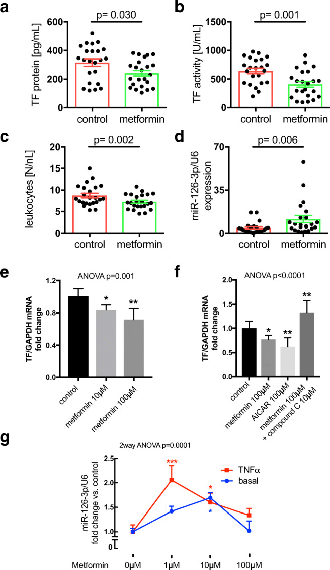
Metformin reduces tissue factor procoagulant activity in diabetes. Patient cohort: Plasma of patients with diabetes receiving metformin or not was analyzed with respect to TF protein (A), TF activity (B), leukocyte count (C), and miR-126 expression (D). n = 46; shown are mean ± SEM; differences between groups were measured by a Student’s t test or Mann–Whitney test. P-values are indicated. In vitro experiments: THP-1 cells were left untreated or incubated with the indicated concentrations of metformin for 24 h and then induced with 10 μg/mL LPS for 2 h and TF mRNA expression assessed (E). THP-1 cells were cultured in the absence or presence of metformin, AICAR, or metformin together with compound C at the indicated concentrations for 24 h. The cells were then stimulated with 10 μg/mL LPS for 2 h and TF mRNA quantified (F). HMEC-1 were left untreated or exposed to different concentrations of metformin as indicated for 72 h. miR-126 expression was then assessed under basal conditions or following stimulation with TNFα for 2 h (G). n ≥ 5; groups were compared by ANOVA with Tukey’s post hoc test (E,F) or 2-way ANOVA with Dunnett’s multiple comparison post hoc test (G). *p < 0.05, **p < 0.01, ***p < 0.0001 vs. control