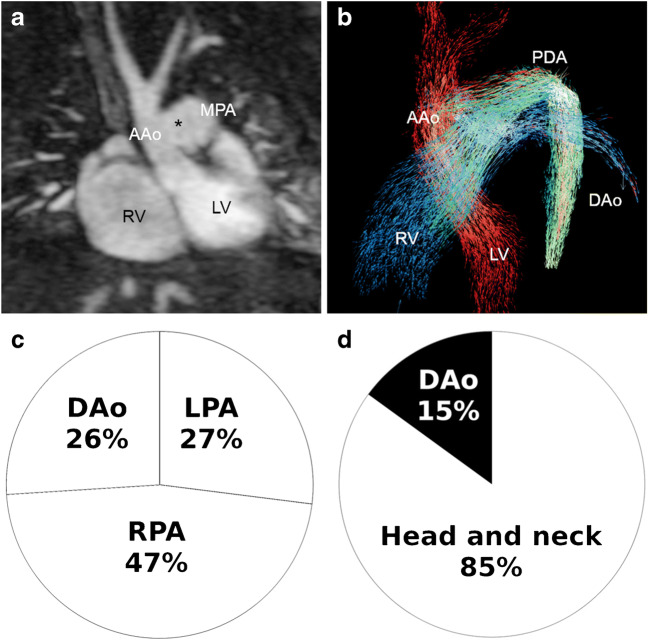
Fig. 10.
Interrupted aortic arch Type A with a large patent duct and aorto-pulmonary window in a 3-day-old girl. a Coronal magnetic resonance (MR) angiography image shows the large aorto-pulmonary window (asterisk) between the ascending aorta (AAo) and the main pulmonary artery (MPA). LV left ventricle, RV right ventricle. b Left oblique 4-D flow vector visualisation reveals mixing of aortic flow (red) and pulmonary flow (blue) through the large aorto-pulmonary window (green) and blood flow via the patent duct (PDA) to the descending aorta (DAo). c, d Four-dimensional flow pathline tracking analysis demonstrates that right ventricular stroke volume is divided into the pulmonary arteries and the DAo somewhat equally (c), whereas most of the left ventricular stroke volume goes into the head and neck vessels and only 15% into the descending aorta (DAo) via the patent duct (d). LPA left pulmonary artery, RPA right pulmonary artery

