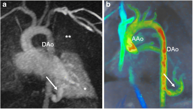
Fig. 12.
Intralobar bronchopulmonary sequestration in the left lower lobe in a 3-month-old boy. a Coronal image of time-resolved magnetic resonance (MR) angiography reveals the systemic artery (arrow) arising from the descending aorta (DAo) at the diaphragmatic level and the delayed pulmonary contrast enhancement (single asterisk) compared with the other pulmonary segments (double asterisks). b Coronal 4-D flow velocity map and flow quantification in the ascending aorta (AAo) and in the systemic artery (arrow) result in a systemic-to-pulmonary shunt of 27% of the cardiac output

