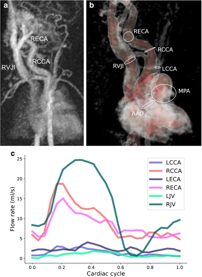
Fig. 13.
Congenital haemangioma presenting as a high-flow lesion in the neck of a 2-day-old boy. a Coronal image of time-resolved magnetic resonance (MR) angiography shows the dilated right common and external carotid arteries (RCCA, RECA) and enlarged internal jugular vein (RVJI) as well as multiple feeding and draining vessels. b, c Corresponding coronal 4-D flow vessel isosurface visualisation (b) and quantification (c) show the asymmetrical flow distribution in the right and left cervical vessels (ovals) because of the high-flow lesion on the right. AAo ascending aorta, LCCA left common carotid artery, LECA left external carotid artery, LVJ left internal jugular vein, MPA main pulmonary artery

