Fig. 4.
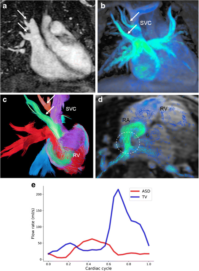
Partial anomalous pulmonary venous connection with superior sinus venosus atrial defect in a 2-year-old boy. Cardiac MRI was performed for assessment of detailed anatomy and measurement of the pulmonary-to-systemic blood flow ratio. There is a haemodynamically relevant left-to-right shunt with a calculated ratio of about 3 based on ventricular stroke volumes. a, b Similar to the coronal magnetic resonance (MR) angiography image (a), coronal 4-D flow velocity map (b) depicts the abnormal drainage of the right upper lobe veins (arrows) into the superior caval vein (SVC). c Right oblique 4-D flow-based color-coded pathline tracking shows that blood originating from the right lower and left pulmonary veins (red) directly passes the atrial septal defect together with blood coming from the abnormal draining right upper pulmonary veins (arrows; orange, green) and the SVC. d, e Axial 4-D flow allows for visualisation of the shunt direction by vectors (d) and direct shunt quantification (dotted circle) as shown in the diagram (e) for the sinus venosus defect and the tricuspid valve. RA right atrium, RV right ventricle

