Fig. 8.
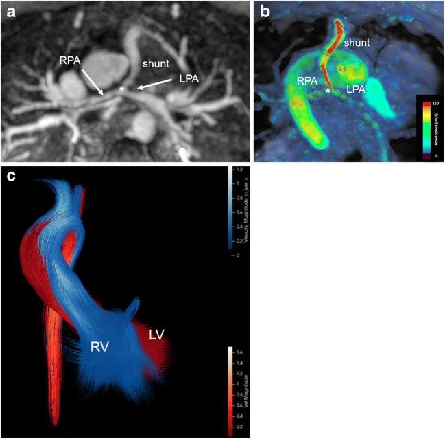
Pulmonary atresia, ventricular septal defect, confluent pulmonary arteries and multiple major aortopulmonary collaterals in an 18-month-old girl after shunt placement between right ventricle and pulmonary artery bifurcation. a Axial magnetic resonance (MR) angiography maximum-intensity projection shows a small-diameter shunt with distal shunt stenosis at the bifurcation (asterisk) as well as a tiny right pulmonary artery (RPA) and a proximally stenotic left pulmonary artery (LPA). b Corresponding axial 4-D flow velocity map reveals high velocities within the shunt (red). Flow assessment is feasible in all three arteries (black ovals). c Coronal 4-D flow colour-coded pathlines demonstrate blood flow separation of the right (RV) and left (LV) ventricles without real mixing despite the ventricular septal defect: RV blood (blue) mainly runs into the brachiocephalic trunk and left carotid artery, whereas LV blood (red) flows into the left subclavian artery

