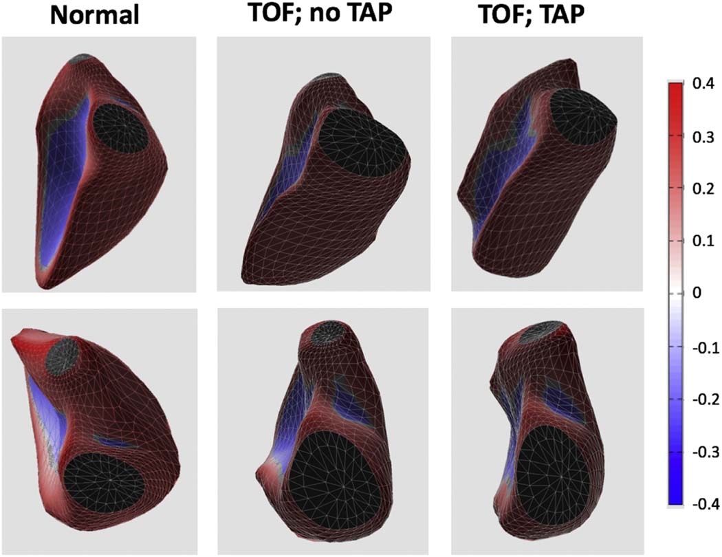
Figure 3.
Examples of 3D casts of the RV cavity obtained in 3 subjects: normal control (left), and two rTOF patients: without (middle) and with (right) transannular patch (TAP). As the mid and apical free wall and the RVOT become more convex (top and bottom, respectively) among these three subjects (left to right), RV curvedness increases in a stepwise manner.

