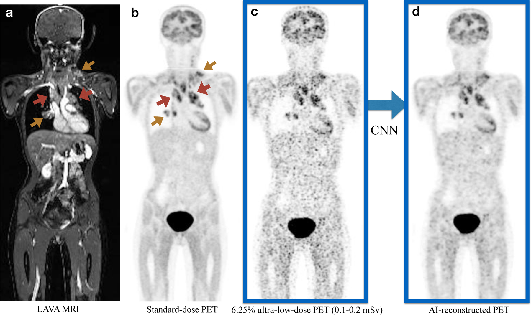
FIGURE 2. Representative 18F-FDG PET/MRI scan of a 16-year old female patient with Hodgkin lymphoma (HL).

a). Coronal contrast-enhanced T1-weighted LAVA (liver acquisition and volume acquisition) MRI; b). Coronal view of a standard dose 18F-FDG dose PET scan (3 mBq/kg); c). Simulated ultra-low-dose PET scan at 6.25% 18F-FDG dose; d). The AI-reconstructed ultra-low-dose 18F-FDG PET image, reconstructed based on the 6.25% ultra-low dose PET and MRI scans as combined inputs. The red arrows point to the hypermetabolic tumors in the mediastinum. Additional hypermetabolic tumors are noted at the right hilum and left lower neck (yellow arrows). All lesions can be detected on all scans, but tumor-to-background contrast and confidence for lesion detection is improved on the AI-reconstructed 18F-FDG PET.
