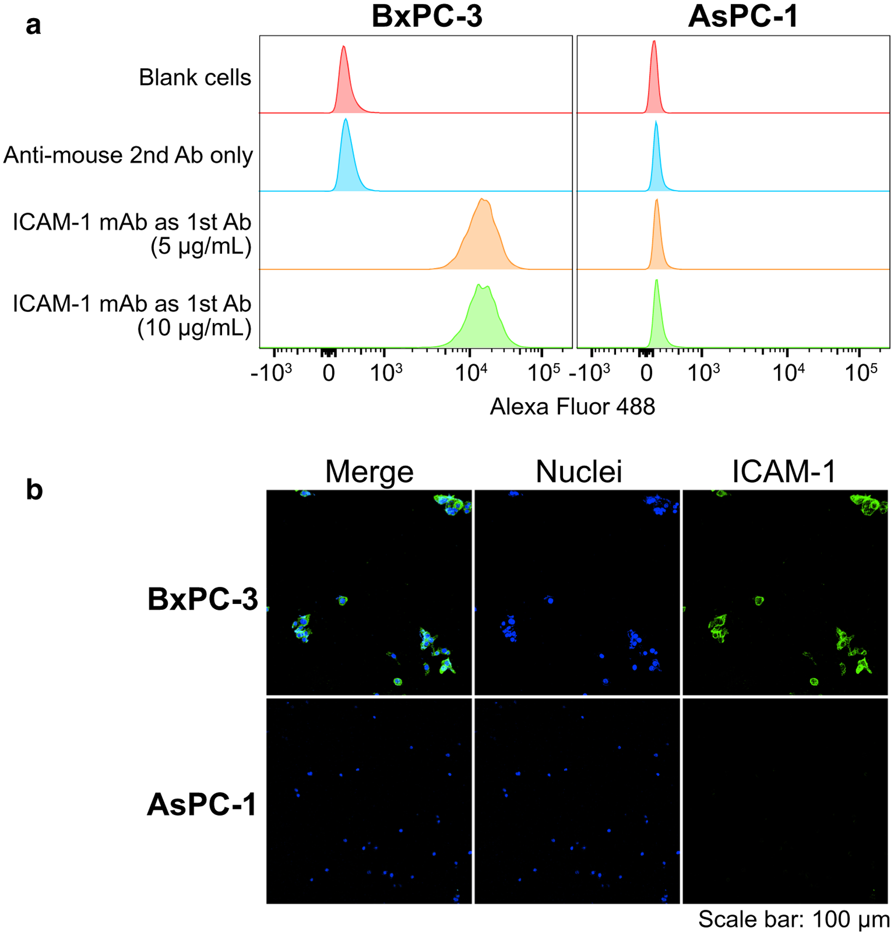
Fig. 1.

In vitro ICAM-1 expression in pancreatic ductal adenocarcinoma (PDAC) cell lines (BxPC-3 and AsPC-1) confocal imaging after immunofluorescent staining. Panels: a flow cytometry (n = 3); b confocal imaging after immunofluorescent staining. Sample groups: the controls were engaging with goat anti-mouse secondary antibody only (anti-mouse 2nd Ab only); the samples were engaging with mouse anti-human ICAM-1 monoclonal antibody as the primary stain (ICAM-1 mAb as 1st Ab); the cell nuclei stained by Hoechst (Nuclei); the samples were engaging with mouse anti-human ICAM-1 monoclonal antibody as the primary stain (ICAM-1)
