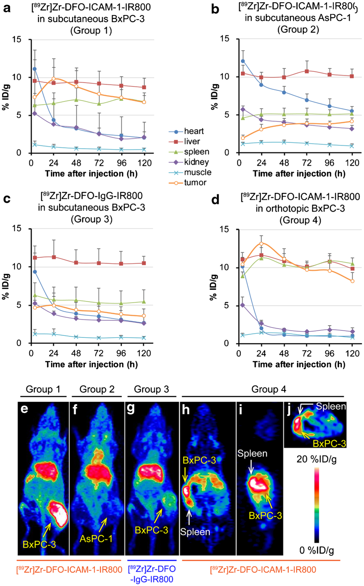
Fig. 2.

The uptake kinetics of region of interest (ROI) and typical maximum intensity projections (MIP) and tomograms at 24 h post-injection (p.i.) in the in vivo positron emission tomography (PET) images of nude mice bearing subcutaneous and orthotopic pancreatic ductal adenocarcinoma (PDAC) tumors (BxPC-3 and AsPC-1). p < 0.05. Panels: a IR800/89Zr dual-labeled ICAM-1 monoclonal antibody ([89Zr]Zr-DFO-ICAM-1-IR800) injected into subcutaneous BxPC-3 models (Group 1; n = 4); b [89Zr]Zr-DFO-ICAM-1-IR800 injected into subcutaneous AsPC-1 models (Group 2; n = 5); c IR800/89Zr dual-labeled IgG isotype control ([89Zr]Zr-DFO-IgG-IR800) injected into subcutaneous BxPC-3 models (Group 3; n = 5); d [89Zr]Zr-DFO-ICAM-1-IR800 injected into orthotopic BxPC-3 models (Group 4; n = 3); e MIP of subcutaneous BxPC-3 models injected with [89Zr]Zr-DFO-ICAM-1-IR800 (Group 1); f MIP of subcutaneous AsPC-1 models injected with [89Zr]Zr-DFO-ICAM-1-IR800 (Group 2); g MIP of subcutaneous BxPC-3 models injected with [89Zr]Zr-DFO-IgG-IR800 (Group 3); coronal (h), sagittal (i), and transverse (j) sections of orthotopic BxPC-3 models injected with [89Zr]Zr-DFO-ICAM-1-IR800 (Group 4)
