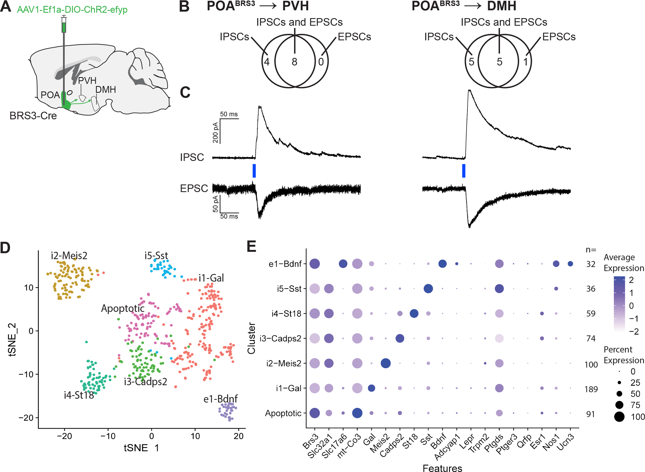
Figure 6.

POABRS3→PVH and POABRS3→DMH neurons are both inhibitory and excitatory and POABRS3 are in multiple clusters. a) Schematic showing injection of Cre-dependent ChR2-EYFP-expressing AAV in the POA and projections to PVH and DMH. b) Venn diagrams showing the number of POABRS3→PVH and POABRS3→DMH neurons with each type of postsynaptic current. EPSC, excitatory postsynaptic current; IPSC, inhibitory postsynaptic current. c) Voltage-clamp trace of POABRS3→PVH and POABRS3→DMH stimulation in PVH- and DMH-containing brain slices. Recordings were made in the presence of tetrodotoxin (500 nM) and 4- aminorpyridine (100 μM), showing monosynaptic inhibitory and excitatory input from the POA to both downstream hypothalamic areas. IPSCs recorded at +10 mV and EPSCs at –55 mV. Traces are mean of 10 – 15 stimulations per cell. d) tSNE plot of BRS3 neuron mRNA expression in the POA region with data from (Moffitt et al., 2018). e) Expression of selected mRNAs in the POA region BRS3 clusters. See also Figure S5.
