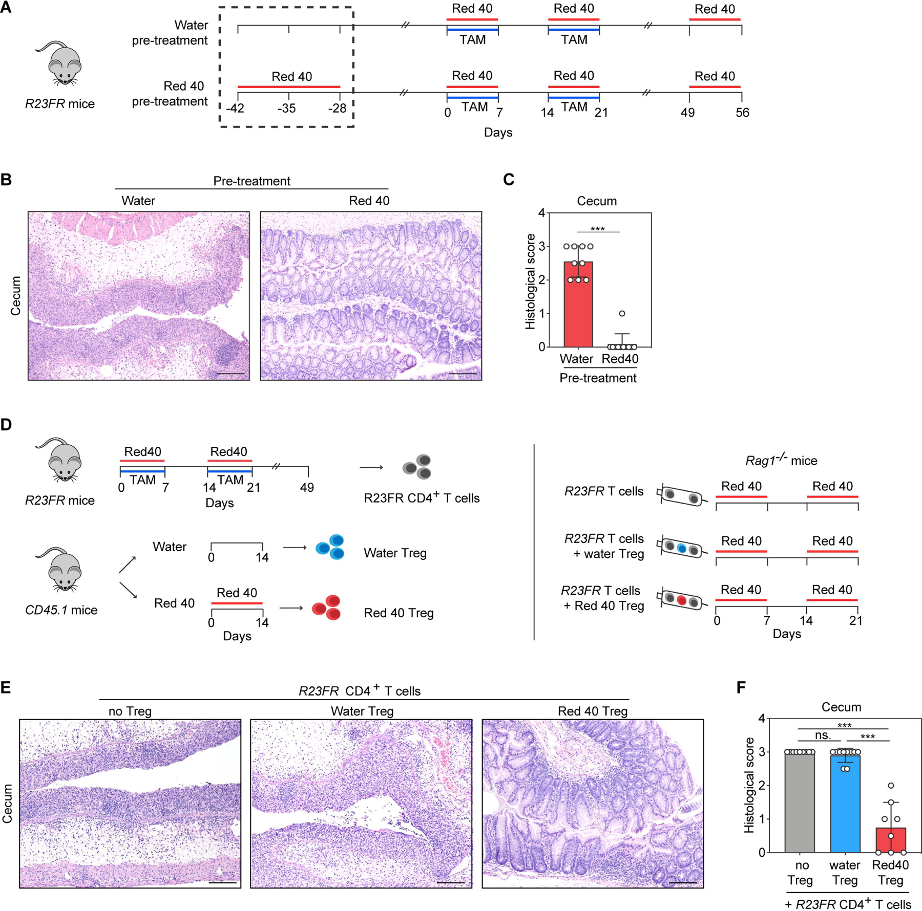
Fig. 2. Red 40 induced Tregs protect from colitis development in R23FR mice.

(A) Experimental scheme. R23FR mice were treated with Red 40 (0.25g/L) for 2 weeks before TAM and Red 40 treatment. (B,C) Representative H&E-stained sections (B) and histologic scores (C) of the cecum of experimental mice at day 56 described in Fig. 2A. (D) Experimental scheme. Unfractionated CD4+ T cells were isolated from the mLN of R23FR mice in remission (d48 after TAM and Red 40 treatment) (106 cells/ mouse) and co-injected with 4× 10⁵ Treg, CD45.1+CD4+CD25+, isolated from WT mice (CD45.1+) treated with or without Red 40 into recipient Rag1−/− mice treated with Red 40 in the drinking water. (E,F) Representative H&E-stained sections (E) and histologic scores (F) of the cecum of experimental mice at d21 described in Fig. 2D. Scale bars in (B) and (E), 50 μm. In (C) and (F), each dot indicates an individual mouse. Error bars indicate SEM. *** p<0.001; ns, p >0.05 by nonparametric Mann-Whitney test.
