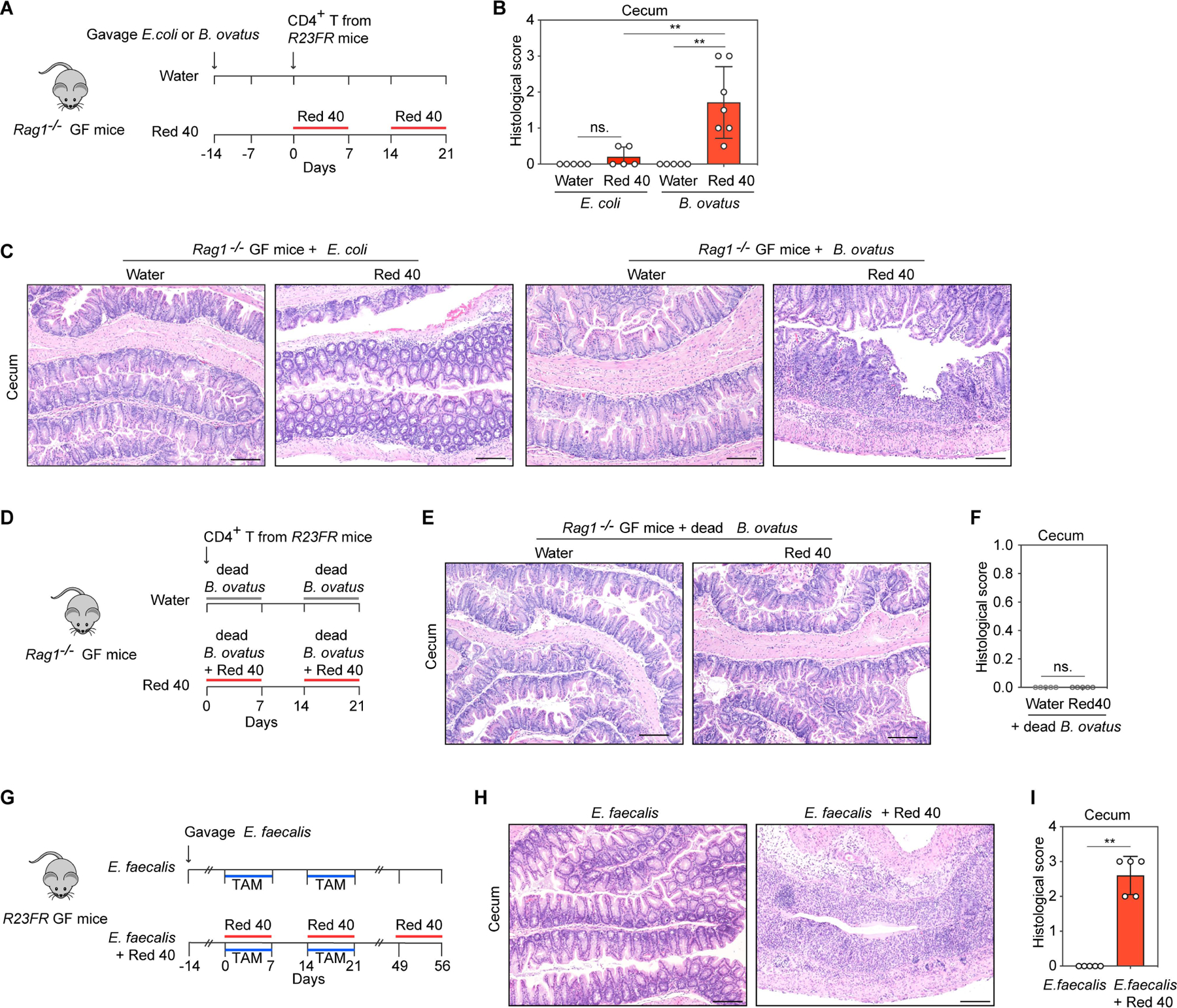
Fig 5. B.ovatus and E.faecalis contribute to Red 40-induced colitis.

(A) Experimental scheme. CD4+ T cells from R23FR mice in remission were transferred into B. ovatus or E.coli monocolonized Rag1−/− germ-free (GF) mice (106 cells/mouse) treated with and without Red 40 in drinking water. (B,C) Histologic scores (B) and representative H&E-stained sections (C) of the cecum of experimental mice at day 21 described in Fig. 5A. (D) Experimental scheme. Rag1−/− GF mice adoptively transferred with CD4+ T cells from R23FR mice in remission (d48 after TAM + Red 40 treatment) (106 cells/mouse) were treated with dead B.ovatus in drinking water (10g/L) or dead B.ovatus (10g/L) plus Red 40 (0.25g/L) in drinking water. (E,F) Representative H&E-stained sections (E) and histologic scores (F) of the cecum of experimental mice at day 21 described in Fig. 5D. (G) Experimental scheme. E.faecalis monocolonized R23FR GF mice were treated with TAM in the food and treated with 0.025% Red 40 (0.25g/L) in drinking water. (H,I) Representative H&E-stained sections (H) and histologic scores (I) of the cecum of experimental mice at day 56 described in Fig. 5G. Scale bars (C), (E) and (H), 50 μm. In (B), (F) and (I) each dot indicates an individual mouse. Error bars indicate SEM. ns, p >0.05, ** p<0.01, by nonparametric Mann-Whitney test.
