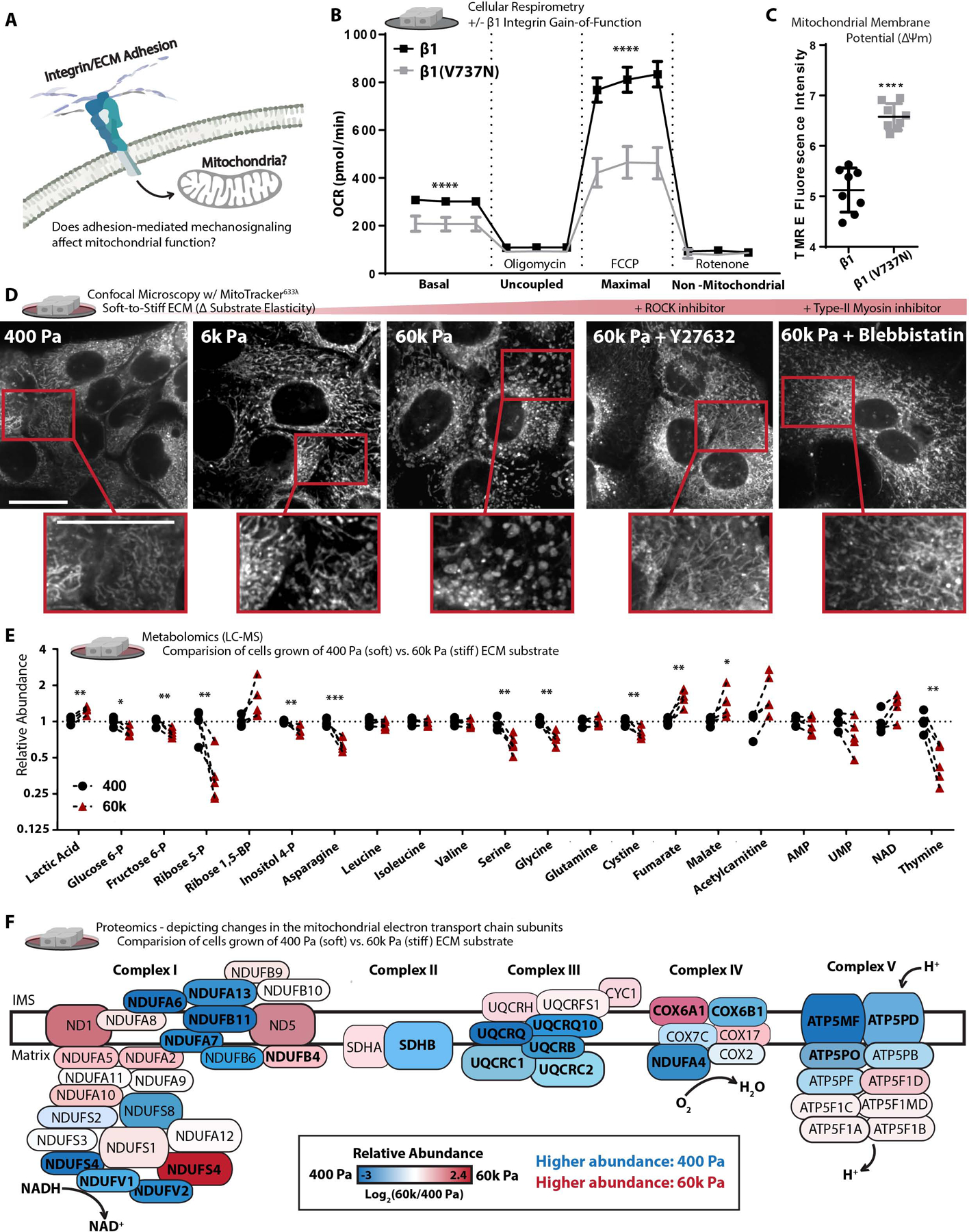
Figure 1. Adhesion-mediated mechanosignaling alters mitochondrial structure and function of human mammary epithelial cells (MECs).

A. Graphical representation of the experimental question.
B. Mitochondrial oxygen consumption rate (OCR) of β1-integrin or β1(V7373N) expressing cells (100k cells per well, n=5 wells, 3 replicate measures, repeated 3 times), mitochondrial stress test conditions: uncoupled = oligomycin [1 μM], maximal = Trifluoromethoxy carbonylcyanide phenylhydrazone (FCCP) [1 μM], non-mitochondrial = antimycin A [1 μM] and rotenone [1 μM].
C. Mitochondrial membrane potential, measured after 1 h treatment of Tetramethylrhodamine ethyl-ester (TMRE) [10 nM] (n = 2 wells, repeated 4 times).
D. Confocal Microscopy depicting mitochondrial network structure in PFA-fixed cells cultured on varied soft-to-stiff fibronectin coated [6 μM/cm2] polyacrylamide hydrogels (soft-to-stiff ECM), for 24 h +/− y27632 [10 μM] or Blebbistatin [10 μM], stained with mitotracker (deep red FM) [100 nM]. (Scale Bar: 10 μm). MitoMAPR Quantification: 400 (18), 6k (20) 60k (7), 60k + Y27632 (15), and 60k + Blebbistatin (12) junctions per network.
E. Selection of metabolites measured with (LC-MS) from MECs cultured on soft of stiff ECM for 24 h, fold change relative to 400 Pa. (n=4–5 biological replicates LC-MS run together, repeated 2 times)
F. Relative abundance (fold change) of mitochondrial ETC subunits measured via timsTOF LC-MS of MECs cultured on soft or stiff ECM for 24 h (n=3 biological replicates). Bolded text indicates *P of < 0.05 or less, locations and sizes of ETC subunits graphically depicted are approximate and not to molecular scale.
