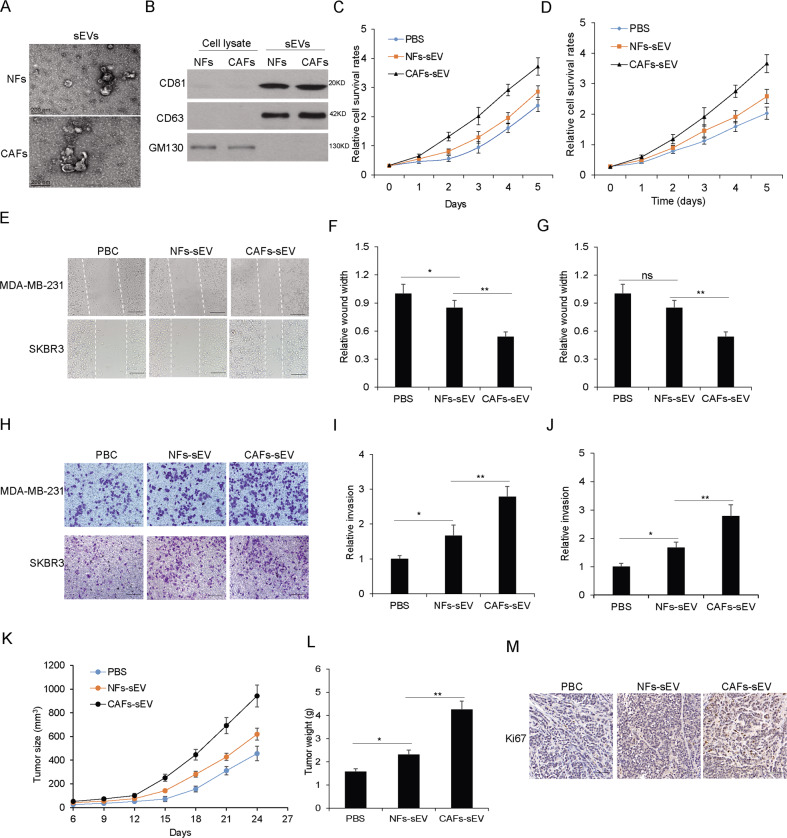
Fig. 1. The sEVs from CAFs increased breast cancer cell survival and metastasis ability.
A The representative picture of sEVs from NFs or CAFs. Transmission electron microscopy was used to take the pictures. B The sEVs markers were identified. The total protein was isolated from NFs or CAFs derived sEVs and performed for CD81, CD63, and GM130 immunoblotting by western blotting. C, D Cell proliferation of MDA-MB-231 and SKBR3 cells exposed to CAFs-sEV and NFs-sEV (25 µg/ml) assayed by CCK8. E Wound healing assay was used to examine cell migration of MDA-MB-231 and SKBR3 cells with CAFs-sEV or NFs-sEV treatment (25ug/ml) (scale bars, 100 µm). F, G Data analysis of (E). H Transwell system was used to examine cell invasion of MDA-MB-231 and SKBR3 cells with CAFs-sEV or NFs-sEV treatment (25 µg/ml) (scale bars, 100 µm). I, J Data analysis of (H). K Tumor growth in vivo. l the average tumor weight. M IHC straining for Ki67 (scale bars, 50 µm). *p < 0.05, **p < 0.01.

