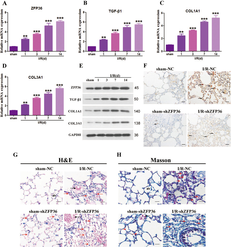
Fig. 6. Role of ZFP36 in intestinal ischemia–reperfusion (I/R)-induced lung fibrosis.
C57BL/6 mice were subjected to 60 min intestine ischemia followed by 60 min reperfusion, and then detections were performed at 1, 3, 7, and 14 days. Sham mice were included as a control. A–D ZFP36 (A), TGF-β1 (B), COL1A1 (C), and COL3A1 (D) mRNA expression in lung tissues of each group were analyzed by RT-qPCR (n = 6 per group, **P < 0.01) and Western blot. E ZFP36, TGF-β1, COL1A1, and COL3A1 protein expression in lung tissues of each group by Western blotting. F Immunohistochemical staining of lung tissues of mice with stable knockdown of ZFP36 or scr for sham and I/R after 14 days. Scale bars: 50 μm. G, H H&E stain (G) and Masson trichrome (H)-stained lung sections. Scale bar: 50 μm.

