Figure 2.
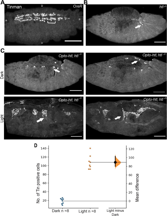
Rescue of htl mutant using Opto-htl. Tin positive heart cells in (A) wild-type and (B) htl homozygous mutant embryo. Tin is expressed in four cardioblasts per hemisegment (brackets) and in a subset of pericardial cells in OreR. Arrows show residual Tin-positive cells in a homozygous htl mutant embryo which fails to develop a heart. (C) twi::Gal4 > Opto-htl is expressed against htl null background and embryos illuminated and stained with Tin antibody. Two homozygous mutants are compared under dark and light conditions for Tin-positive cells. Arrows indicate Tin positive cells of the heart specified on the dorsal side of the embryos. (D) Quantification of the number of Tin-positive cells of the heart at stage 16 under dark and light conditions for embryos described in (C). Scale bar = 50 mm. A = Anterior, P = Posterior, D = Dorsal, V = Ventral view. In (D), the black bar on the right represents the 95% confidence interval with the Bootstrap distribution shown in orange69.

