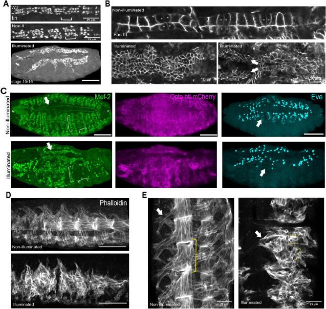
Figure 3.
Mesoderm specific defects in twi::Gal4 > UAS—htl-CRY2-mCherry embryos kept under constant light. (A) Tinman staining pattern in illuminated wild-type (WT) embryos, twi::Gal4 > Opto-htl embryos kept under dark and illuminated conditions. (B) twi::Gal4 > Opto-htl embryos stained with Fas3 and imaged at stage 16 under dark conditions (top) and when illuminated by constant 488 nm light (bottom). Svp-positive cells, marked with an asterisk, are identified by lower Fas3 expression. Arrows denote branching of the heart structure. (C) twi::Gal4 > Opto-htl embryos stained with Mef-2 and Eve antibodies at stage 16 under dark and illumination conditions. Arrows correspond to phenotypes described in the text. (D) Muscle structure in twi::Gal4 > Opto-htl embryos kept under dark and light conditions visualised using Phalloidin staining. (E) As (D) with muscles imaged at higher magnification. Arrows point to extended Ventral Oblique muscles, brackets represent extended muscle fibres, square box shows unfused myoblasts. Scale bar = 50 μm unless stated otherwise. VM = Visceral mesoderm, CB = Cardioblasts, PC = Pericardial cells.

