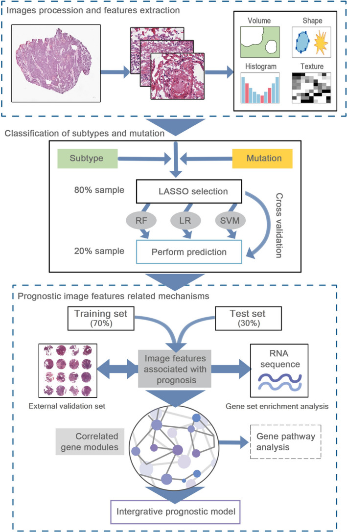
FIGURE 1.

The workflow of data analysis and integration. First, we performed the histopathological image processing and feature extraction. Secondly, three classifiers were constructed by feature selection and 5‐fold cross‐validation, and applied to classify the somatic mutations, transcriptional, and methylation subtypes. Subsequently, we selected the prognostic image features, used bioinformatics analyses to identify correlated gene modules, and established an integrative prognostic model to improve prognosis prediction
