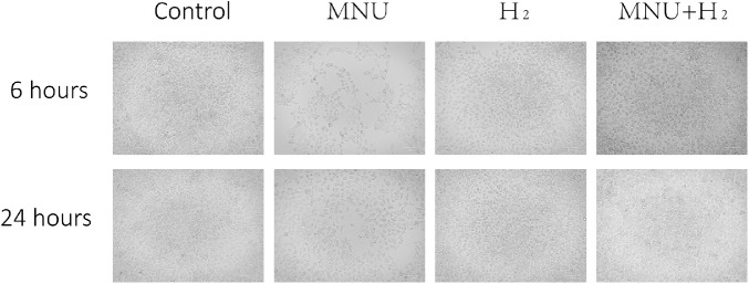
Figure 7.
Images of CECs collected at 6 hours and 24 hours. Cells in the control group adhered to the wall, polygonal, and uniform in size. After co-culture with MNU for 6 hours and 24 hours, the CECs were found significantly sparse than those in the control group. CECs in the MNU group were no longer polygonal in shape. The CECs of the MNU + H2 group were obviously protected by H2 from the killing effect of MNU, although the cell morphology was still very different from that of the control group. Scale bar = 100 µm.

