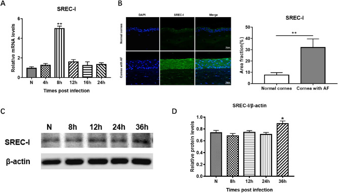
Figure 1.
Expression of SREC-Ⅰ in normal and A fumigatus–treated HCECs and corneas. SREC-Ⅰ mRNA level was significantly higher in A fumigatus–stimulated HCECs at 8 hours comparing with control (A). Immunofluorescence staining for SREC-Ⅰ (green) in normal and A fumigatus–infected human corneas, and quantitative analysis (n = 6/group/time) (t-test). Scale bar = 50 µm. (B) SREC-Ⅰ protein expression level in A fumigatus–stimulated HCECs at 0, 8, 12, 24 and 36 hours (C) and quantitative analysis (D). Values represents means ± SD. *P < 0.05, **P < 0.001 compared with untreated cells by one-way ANOVA test.

