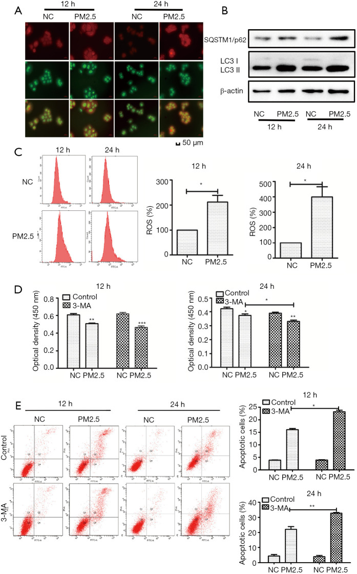
Figure 2.
Autophagy plays a protective role in HaCaT. (A) AO staining results depict a significant accumulation of autophagosomes in HaCaT exposed to PM2.5. (B) Autophagy-related proteins were detected by western blot. (C) ROS generation was assessed by the H2DCFDA assay at 12 and 24 h. Cells were pre-treated with 3-MA for 2 h, cell viability (D) was detected by CCK-8, and apoptosis (E) was detected by Annexin V-FITC/PI double staining and flow cytometry. Magnification: 200×. Each experiment was performed in triplicate and data are presented as mean ± SD. One-way ANOVA and Dunnett’s Multiple comparison test were used to analyze the data (*P<0.05, **P<0.01).

