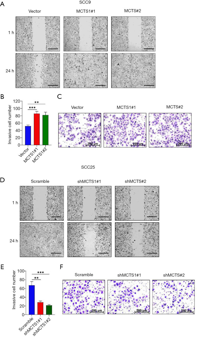
Figure 3.
Cell migration and invasion assay of MCTS1-konckdown and overexpressed oral cancer cells evaluated by wound healing and Transwell invasion tests. (A) Wound healing of MCTS1-overexpressed SCC9 cells at 1 hour and 24 hours (scale bar =500 µm). (B) Invasive cell number of MCTS1-overexpressed SCC9 across the Transwell membrane. (C) Representative migrated cells images for SCC9 cells. (D) Wound healing of MCTS1-knockdown SCC255 cells at 1 hour and 24 hours (scale bar =500 µm). (E) Invasive cell number of MCTS1-knockdown SCC25 cells across the Transwell membrane. (F) Representative migrated cells images for the SCC25 line. MCTS1, malignant T-cell-amplified sequence 1. Statistic analysis: Two-tail Student’s t-test, **P<0.01, ***P<0.001, significantly different from vector or scramble group.

