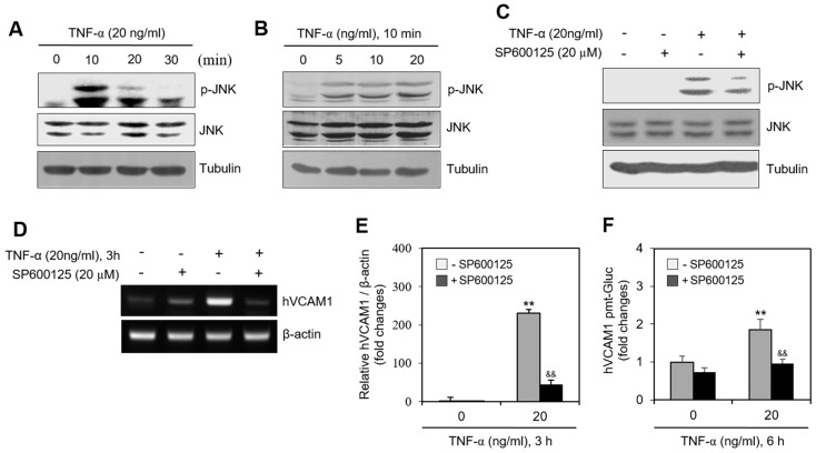
Figure 5.
JNK activation by TNF-α regulated human vascular cell adhesion molecule-1 (hVCAM1) expression. (A,B) MH7A cells were treated with 20 ng/mL TNF-α for various times (A). MH7A cells were treated with various concentrations of 20 ng/mL TNF-α for 10 min (B). JNK and phosphorylated (p)-JNK were measured by western blot analysis. (C,D) MH7A cells were treated with 20 ng/mL TNF-α in the presence or absence of SP600125, JNK inhibitor. were used for inhibition of JNK activation and 20 μM of SP600125 had strong ability of inhibition of JNK activation. Cell lysates were prepared and protein level of JNK and p-JNK was detected by western blot analysis (C). RNA was purified with Nucleozol®. hVCAM1 and hBAFF expression was detected by RT-PCR (D). (E) MH7A cells were transfected with pEZx-PG02-hVCAM1-gaussia luciferase (Gluc) plasmid DNA by using polyethylenimine (PEI), and treated with TNF-α in the presence or absence of SP600125. Gluc activity was measured by using luminometer. (F) MH7A cells were treated with 20 ng/mL TNF-α for 6 h in the presence or absence of SP600125. Then, WiL2-NS cells were added and co-incubated to adhere for 30 min. Unbound WiL2-NS B cells were washed out and MTT assay was used to analyze bound B cells. Each experiment was performed at least four times. Data in a bar graph represent the means ± SD. ** p < 0.01; significantly different from SP600125-untreated and TNF-α-untreated control group. && p < 0.01; significantly different from SP600125-untreated and TNF-α-treated control group (E,F).

