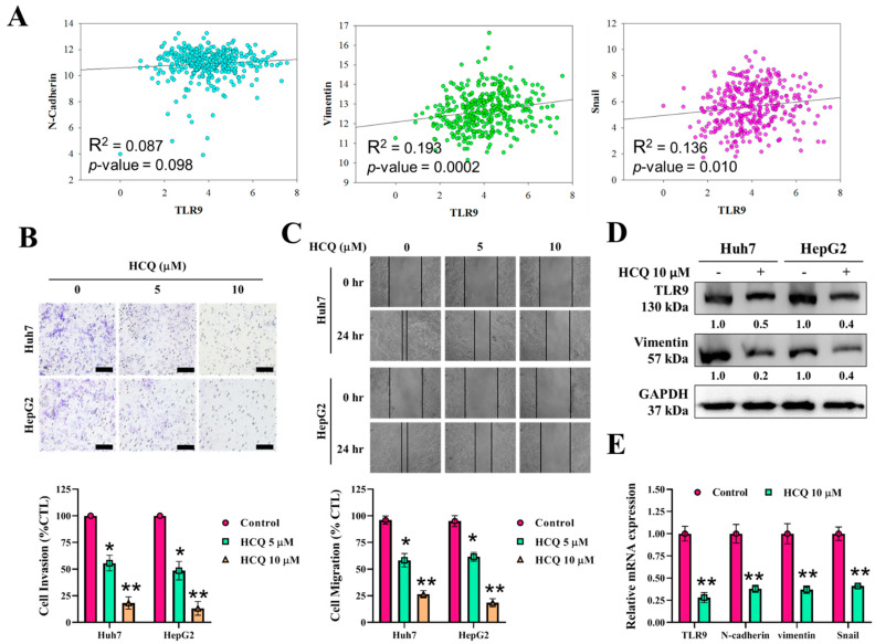
Figure 4.
HCQ inhibited HCC cancer cells’ oncogenic and invasive properties. (A) Correlation analysis of TLR9 expression with EMT associate marker expression. (B,C) HCQ significantly inhibited HCC (Huh7 and HepG2) cell invasive and migratory abilities. The representative bar plot represents the quantification of invasion and migratory abilities after HCQ treatment calculated by ImageJ, provided in (B) (below) and (C) (below), respectively. (D) Western blot analysis of expression of TLR9, and EMT marker (vimentin); GAPDH was used as an internal control. Representative protein bands are shown a reduction in expression of TLR9 and vimentin observed after the HCQ treatment (10 μM). (E) The mRNA expression level of TLR9 and EMT markers (N-cadherin, vimentin, and Snail2) were determined using real-time qRT-PCR; GAPDH was used to normalize the expression levels. Data are the mean ± SEM of three independent experiments. * p < 0.05, ** p < 0.01.

