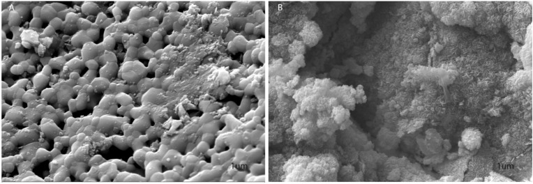
Figure 1.
Scanning electron microscopy (SEM) analysis of S-HA and B-HA. (A) SEM S-HA images show its porous structure, Scale bar: 1 μm, × 11,57 K. (B) B-HA structure is characterized by nano-metric particles forming thin lamellae with a microstructure composed of nano-size building blocks and multi-scale porosity, Scale bar: 1 μm, × 10,210 K.

