Figure 1.
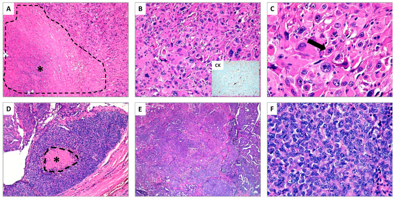
(A–C) Anaplastic thyroid carcinoma (ATC) (hematoxylin and eosin stain); (A) The presence of extensive tumor necrosis is a typical aspect of ATC (see asterisk *) (original magnification ×10); (B) The neoplastic cells show marked nuclear atypia with spindled and pleomorphic morphology, associated to elevated mitotic rate, simulating high-grade pleomorphic sarcoma. In the insert, focal immunostaining for cytokeratins supports the epithelial nature of ATC (original magnification ×20); (C) At higher magnification, the pronounced nuclear atypia and an atypical mitosis (see arrow) are shown (original magnification ×40). (D–F) Poorly differentiated thyroid carcinoma (PDTC) (hematoxylin and eosin stain); (D) The typical example of PDTC is the so-called “insular carcinoma”. In this field, the tumor shows a small focus of tumor necrosis (see asterisk *) (original magnification ×10); (E) The neoplasm exhibits a prevalent solid growth pattern (original magnification ×20); (F) At higher magnification, the tumor cells appear small and uniform, the nuclei are generally rounded and hyperchromatic, in absence of the typical aspects of papillary thyroid carcinoma (original magnification ×40).

