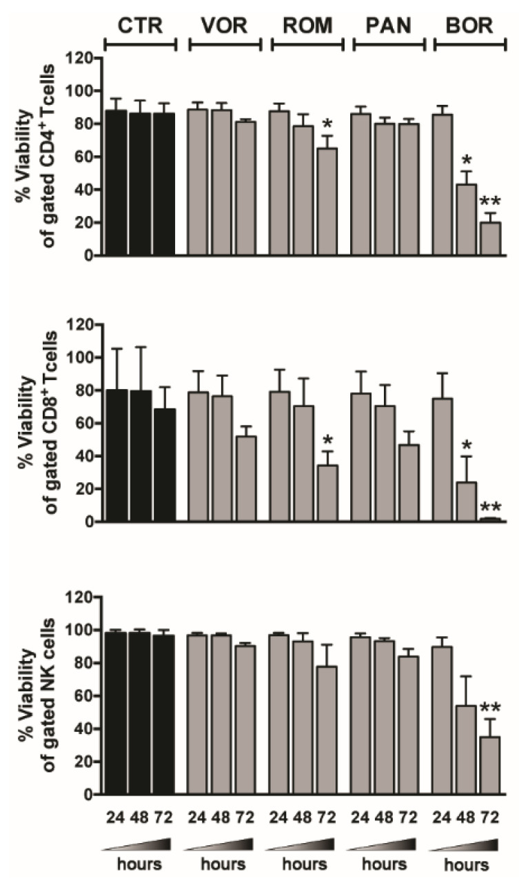
Figure 2.
Lymphocyte viability in LRA-treated PBMCs. PBMCs were cultured in medium alone (control, CTR) or supplemented with 334 nM VOR, 10 nM ROM, 20 nM PAN, 5 nM BOR; after 24, 48, and 72 h, the viability of CD4+ T (top), CD8+ T (center), and NK cells (bottom) was examined by LIVE/DEAD staining and flow cytometry analysis of gated CD3+CD4+, CD3+CD8+, and CD3−CD56+CD16+/− cells, respectively. Bars represent mean ± SEM (n = 4). * p < 0.05, ** p < 0.01 by paired t-test.

