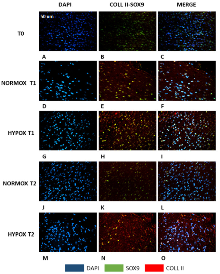
Figure 2.
Double immunofluorescence of meniscal samples. A–C: native meniscus; D–F: meniscus cultured under normoxia for 7 days (T1); G–I: meniscus cultured under hypoxia for 7 days (T1); J–L: meniscus cultured under normoxia for 14 days (T2); M–O: meniscus cultured under hypoxia for 14 days (T2). Blue: DAPI; green: SOX-9; red: collagen type II; yellow: co-expression of SOX-9 and collagen type II. Scale bar for all images: 50 µm.

