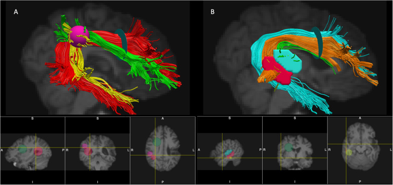
FIGURE 1.
Placement of ROIs and segmentation of the AF (the right hemisphere is shown). Panel (A) - segmentation according to the modified Catani model: AF long – red, AF anterior – green, and AF posterior – yellow. Panel (B) – segmentation according to the modified Glasser and Rilling model: AF-STG – green, AF-MTG- orange, and AF Temporal Pole – cyan. The ROIs used for segmentations of the tracts: Frontal ROI – light green 2D disk, Temporal ROI – red 2D disk, Parietal ROI – pink 3D sphere, STG ROI – cyan manually drawn region, MTG ROI – red manually drawn region, Temporal Pole ROI – yellow 2D disk (see text for more details per reconstruction criteria). Note that 2D disks used as ROIs (Frontal ROI, Temporal ROI, Temporal Pole ROI) are visualized in TrackVis as 2D spheres on the bottom panels.

