FIGURE 1.
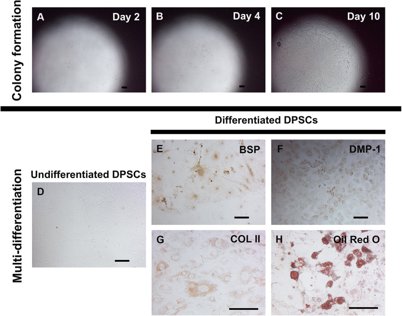
Self-renewal and in vitro multi-differentiation of murine DPSCs. Murine DPSCs were isolated from dental pulp of neonatal lower molar teeth. A low density of cells (1000 cells/cm2) were plated for several single cell depositions to observe a colony forming ability and characterize for their multi-differentiation. Representative figures show at the same spot of DPSC clone at different time points, Day 2 (A), Day 4 (B), and Day 10 (C). The undifferentiated DPSC clone proliferated to gain the larger size, indicating its self-renewal property. The in vitro differentiation in various specific differentiation media demonstrates DPSC multi-differentiation potential. Undifferentiated DPSCs cultured in the stem cell media were spindle-shaped, indicating mesenchymal-like stem cell morphology (D). Differentiated DPSCs cultured in the osteo-odontogenic media exhibited positive anti-BSP (in brown) (E) and anti-DMP-1 (in brown) (F) staining. Cultured in the chondrogenic media, differentiated DPSCs showed positive anti-COL II (in brown) (G) staining. Strongly positive Oil Red O staining (in red) (H) was seen in DPSC-derived lipid-containing cells in the adipogenic media. Scale bars indicate 100 μm.

