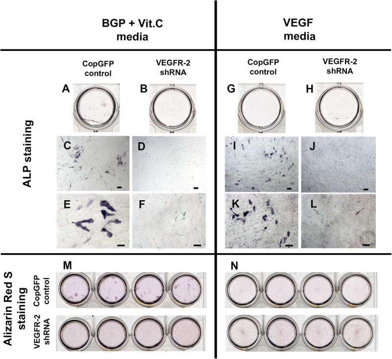
FIGURE 5.
Alkaline phosphatase and Alizarin Red S staining in CopGFP control and VEGFR-2 shRNA DPSCs after a culture in BGP + Vit. C media and VEGF media. After induction in BGP plus vitamin C, some differentiated CopGFP DPSCs (A,C,E) but not VEGFR-2 shRNA DPSCs (B,D,F) demonstrated positive staining of alkaline phosphatase. Alizarin Red S staining showed some positive mineralized nodules in CopGFP DPSCs but not VEGFR-2 shRNA DPSCs (M). For VEGF induction, a similar results of alkaline phosphatase staining were observed in both CopGFP control (G,I,K) and VEGFR-2 shRNA DPSCs (H,J,L), compared to the BGP plus vitamin C. Interestingly, no mineralized nodule was found in both cell groups after VEGF induction (N). Scale bars indicate 100 μm.

