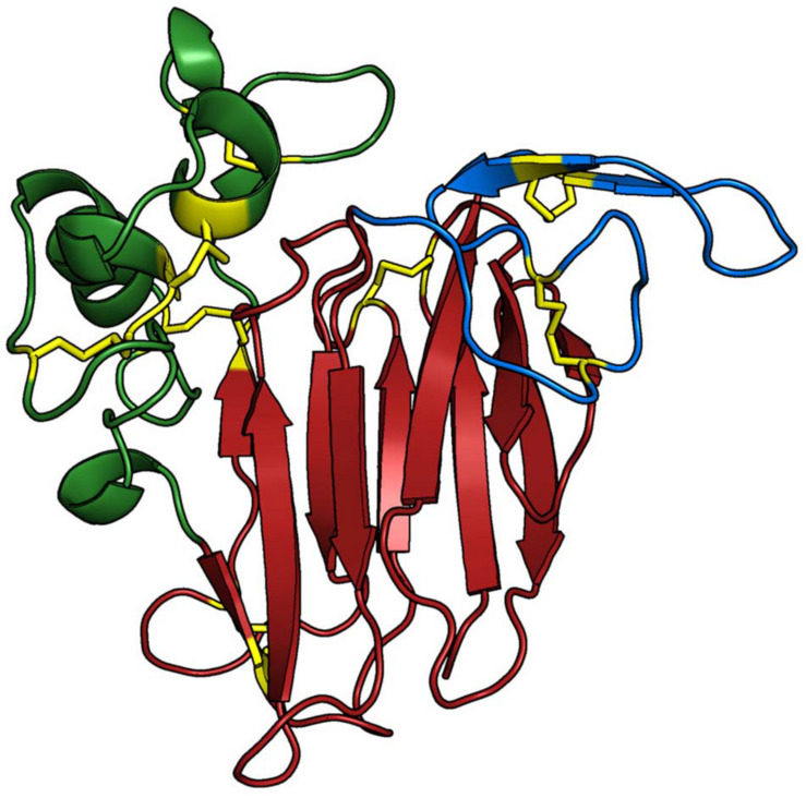
FIGURE 3.
Three-dimensional structure of Gma_P21. The protein structure is coloured based on its domains: (i) domain I (red), consisting of a 11 stranded β-sheet organised as a β-barrel that forms the protein core; (ii) domain II (green), consisting of an α-helix and a set of disulphide-rich loops; and (iii) domain III (blue), presenting a β-hairpin and a coil region, which are both maintained by one disulphide bond each. The main chain of the Osmotin structure is represented as cartoon and the side-chains of the residues involved in disulfide bonds are presented as yellow sticks. The Sni_SnOLP, Sni_SindOLP, Sni_Jami, Gma_OLPb, Gma_P21like, Gma_P21, Gma_OLPc, and Gma_OLPb structures share the same topology. The image was generated with PyMOL Molecular Graphics System version 1.5.0.4 (Schrödinger, LLC).

