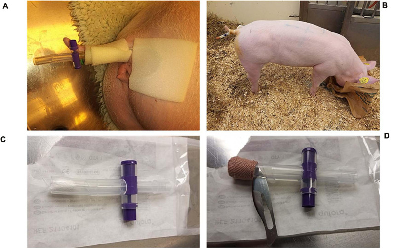
FIGURE 2.
Catheter fixation. (A) External fixation of the catheter. (B) The catheters were easily accessible, and due to proper training prior to the experiment, urine samples could be collected without the need for sedation. (C) To limit contamination of the catheters from the environment, the catheter end was always covered with a replaceable catheter valve that was either closed when accumulating urine for sampling (A) or open and sealed with gauze swaps (C) and secured with tape (D) so that urine could slowly drip through. (D) A secondary non-return valve was made by cutting a thin slit in the silicone right above the gauze. The catheter valve was always discarded before urine sampling and replaced with a sterile valve after sampling.

