
Figure 1.
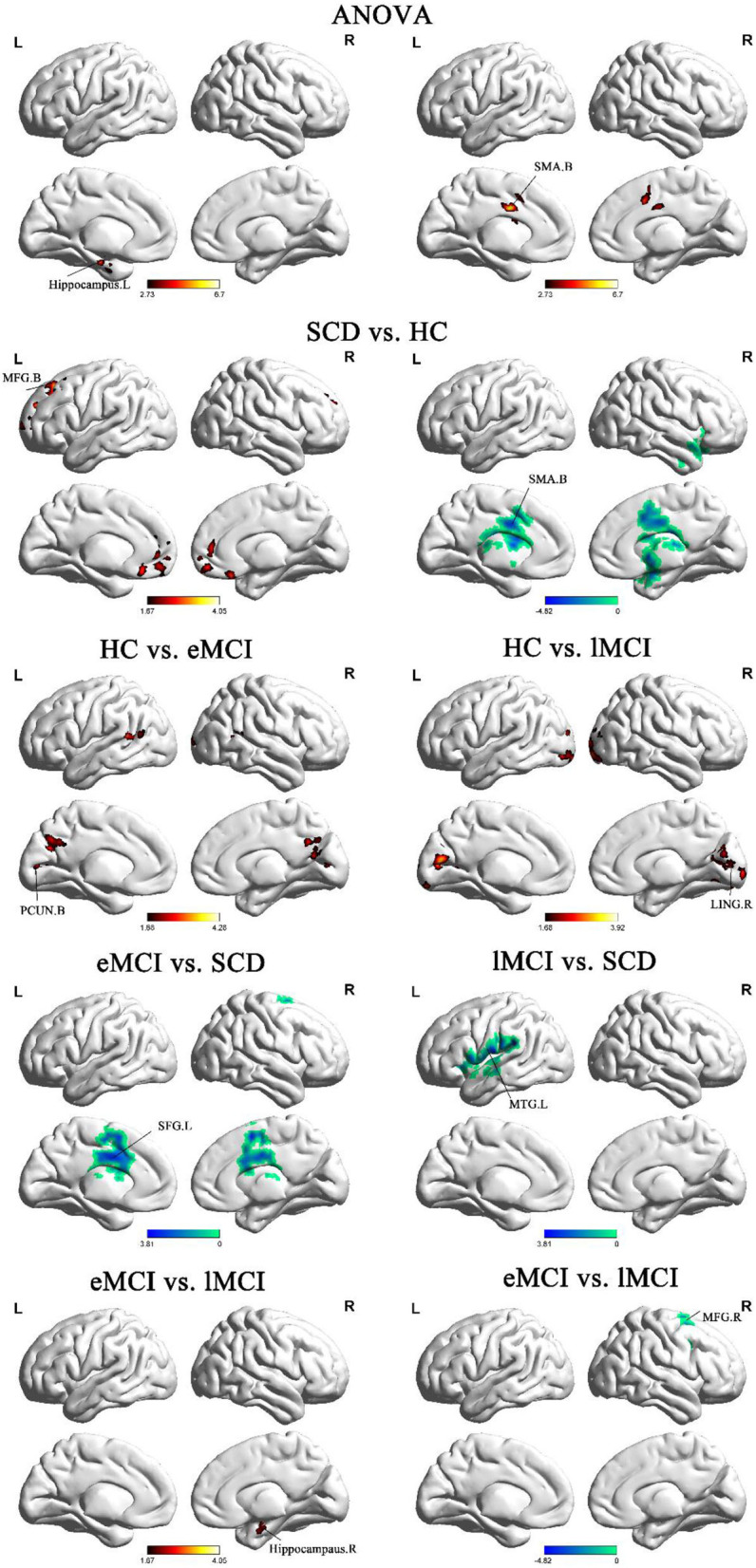
Brain regions exhibiting significant differences in BNM-FC based on analysis of variance (ANOVA) analysis and two-sample t-tests. Age, gender, and years of education were used as covariates for all these results. GRF corrected, cluster size ≥50 mm3, p < 0.05. eMCI, early mild cognitive impairment; lMCI, late mild cognitive impairment; SCD, subjective cognitive decline; HC, healthy controls; SMA, supplementary motor area; PCUN, precuneus; SFG, superior frontal gyrus; MFG, middle frontal gyrus; MTG, middle temporal gyrus; LING, lingual gyrus; L, left hemisphere; R, right hemisphere; B, bilateral hemisphere.