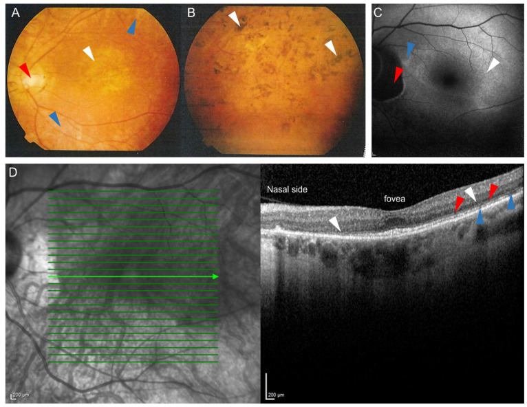
Figure 2.
Representative fundus photographs (A,B), fundus autofluorescence image (C) and an optical coherence tomography (OCT) scan (D) of patients carrying the c.12delC mutation in LRAT suffering from LRAT-associated retinal dystrophy. Fundus photograph of the central (A) and peripheral area (B) of the left eye of a 39-year old patient. (A): Atrophic alterations of the retinal pigment epithelium in the macula (white arrow) and around the vascular arcades (blue arrows), along with retinal atrophy around the optic disc, which shows some temporal pallor (red arrow) are visible. The vessels are attenuated. (B): The peripheral retina showed retinal atrophy and bone-spicule-like hyperpigmentation. (C): Fundus autofluorescence image of a 57-year old patient, showing a subtle hyperautofluorescent ring around a relatively preserved central macula (white arrow) and a juxtapapillary patch of absent autofluorescence (red arrow), sharply outlined by a hyperautofluorescent border. The inferior posterior pole shows granular hypo-autofluorescence (blue arrow), indicating more atrophy of the retinal pigment epithelium in the area outside of the hyperautofluorescent ring. (D): Spectral-domain OCT (SD-OCT) scan of a 57-year old patient, showing relative preservation of the outer nuclear layers, the external limiting membrane, and the ellipsoid zone at the level of the fovea. In the parafovea and perifovea, thinning of the outer nuclear layer is seen (white arrows), along with interruptions of the external limiting membrane (red arrows) and the ellipsoid zone (blue arrows). These interruptions, along with the outer nuclear layer thinning, increase towards the peripheral macula and are more profound on the nasal side.

