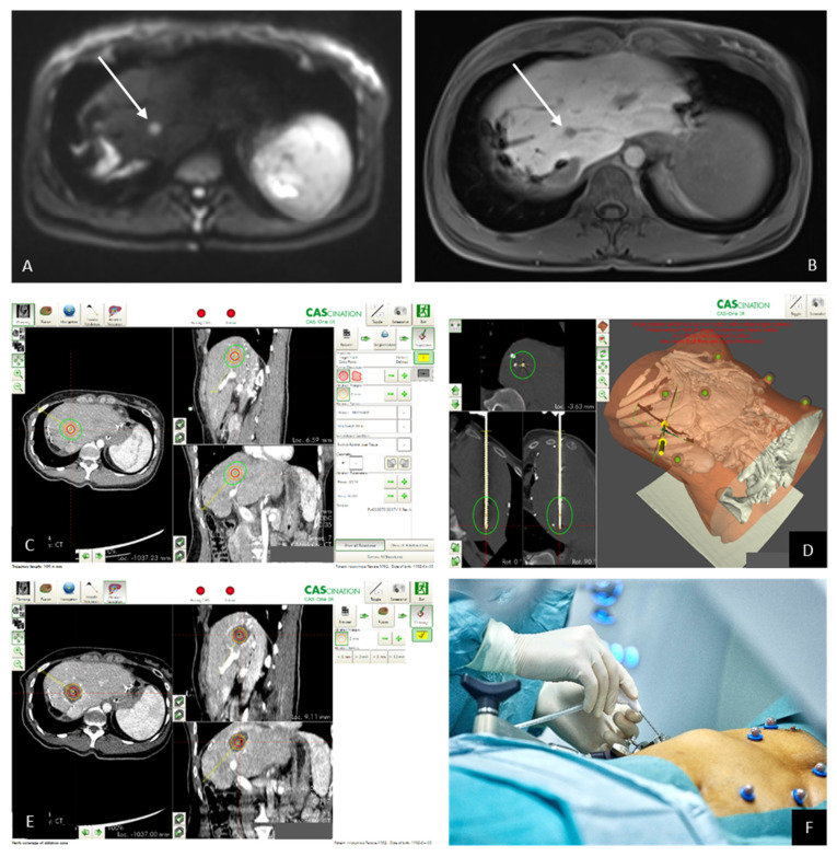
Figure 2.
(A) Pre-interventional MRI imaging reveals the metastasis with high signal in the b800 image of the diffusion-weighted imaging (DWI) and also shows (B) a lack of intracellular uptake of hepatocyte-specific contrast medium in the hepatobiliary phase. (C) The pre-interventional planning of the ablation zone (red circle = segmented tumor; (D) green circle = anticipated ablation zone) needle validation scan with the needle in place and the anticipated ablation zone in green. (E) The ablation zone in the contrast-enhanced control scan; (F) needle in place.

