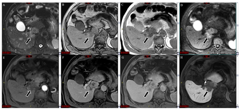
Figure 1.
MRI with Gd-EOB-DTPA of a histologically confirmed intra-hepatic cholangiocarcinoma located in the segment I of the liver (arrow). The lesion showed hyperintensity at the T2-weighted image (A), whereas it was hypo both in in- and out-of-phase T1-weighted images (B,C), coupled with strong hyperintensity in the diffusion-weighted image (D). No significant hyperenhancement was seen in the arterial phase (E). A heterogeneous centripetal enhancement was detected during the portal and late venous phases (F,G), without any washout, but the center of the lesion was continuously hypointense. In the hepatobiliary phase, the lesion appeared hypointense (H).

