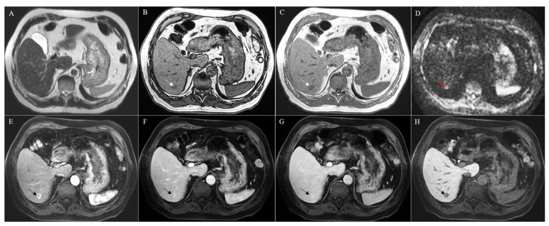
Figure 2.
MRI with Gd-EOB-DTPA of a histologically confirmed combined hepatocellular cholangiocarcinoma located in the segment VI of the liver (arrow). The lesion presented without strong hyperintensity at the T2-weighted image (A) but showed hypointensity both in in- and out-of-phase T1-weighted images (B,C) and strong hyperintensity in the diffusion-weighted image (D). A strong hyperenhancement was evident during the arterial phase (E), followed by a persistent enhancement during the portal and late venous phases (F,G). In the hepatobiliary phase, the lesion appeared hypointense (H).

