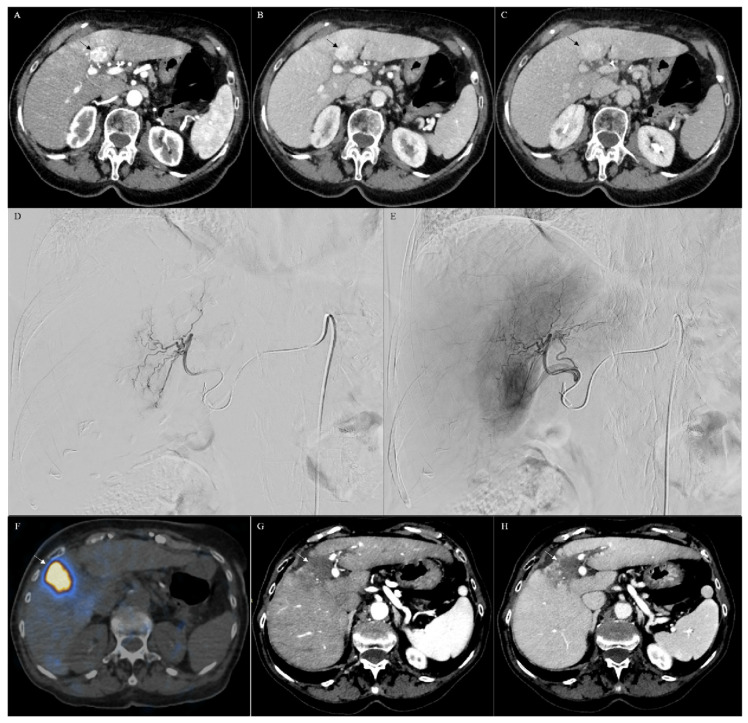
Figure 5.
CT of a combined hepatocellular cholangiocarcinoma (arrow) (A–C). In particular, CT showed a lesion in the segment IV of the liver with a strong hyperenhancement during the arterial phase (A), followed by a persistent enhancement during the portal (B) and delayed phases (C), consistent with the imaging diagnosis of combined hepatocellular cholangiocarcinoma, thereafter confirmed with biopsy. The lesion was treated by using transarterial radioembolization (TARE). In particular, during the angiographic study (D,E), the artery feeding the liver segment IV was selectively catheterized (D), the lesion was confirmed with selective angiography (E), and the intra-arterial treatment was selectively performed. Positron emission/computerized tomography (PET/CT (F)) demonstrated that the treatment was correctly performed, covering all the lesion (arrow in (F)). CT scan, performed 6 months after TARE, demonstrated complete response of the lesion characterized by no vascularized area in the IV segment of the liver in the arterial (arrow in (G)) and in the venosus (arrow in (H)) phases.

