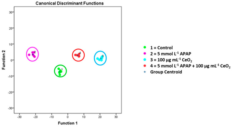
Figure 7.
Metabolic changes of cell membrane lipid patterns of HuH-7 cells after treatment with APAP (5 mmol L−1), CeO2 NPs (100 µg/mL) and APAP (5 mmol L−1) plus CeO2 NPs (100 µg/mL), as identified by means of ToF-SIMS in combination with multivariate data analysis. The diagram shows the values of the discriminant scores obtained from Fisher’s discriminant analysis of 24 HuH-7 samples. The model was evaluated using the “leave-one-out” formalism (100% correct grouping of ungrouped cases).

