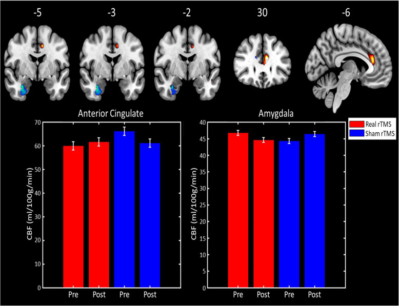
Fig. 2.
Top: Brain regions where regional CBF demonstrated a treatment-by-time interaction. Regions in blue have an interaction driven by treatment-related decreases in CBF following real rTMS, whereas those in yellow/red are driven by CBF reduction in the sham rTMS group. Brain images shown at an uncorrected threshold of p < 0.005, slice numbers are in MNI space. Bottom: Bar graphs showing the cluster mean regional CBF values for the anterior cingulate cluster (left) and right amygdala (right) for the real rTMS (in red) and sham rTMS (in blue) groups

