Figure 1.
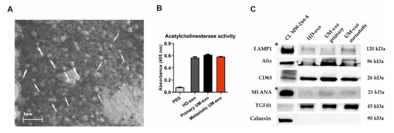
The characteristics of exosomes isolated from the serum of uveal melanoma patients with primary and metastatic disease (UM-exo primary and UM-exo metastatic) and healthy donors (HD-exo). The scanning electron microscopy (SEM) showed the proper shape and size (exosomes marked with white arrows) (A). The isolated exosomes indicate high levels of acetylcholinesterase activity. Data represent the mean ± SD from three technical replicates of absorbance readout at 405 nm from the pooled serum (B). The expression of proteins characteristic for small extracellular vesicles—endosomal origin (LAMP1, Alix, CD63), their protein cargo (TGFβ1), the tumour-origin (MLANA), and absence of organelle-specific proteins (Calnexin) was confirmed by Western blot. The band marked with * were obtained from the short-time exposure of the same membrane, due to differences in the protein expression level between the cell line lysate and exosomes (C). The uncropped Western Blot images can be found in Figure S1.

