Figure 2.
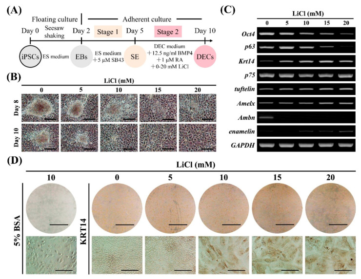
Effects of LiCl concentration on dental epithelial cell (DEC) induction. (A) Diagram of DEC induction from Amelx-iPSCs. After surface ectoderm (SE) induction (stage 1) by SB43 (nodal signaling inhibitor), the cells were treated with BMP4, RA, and LiCl (Wnt/β-catenin pathway activator: 0–20 mM) in DEC medium for DEC induction (stage 2). (B) Cell morphology at days 8 and 10 during stage 2 of induction. Scale bars: 200 μm. (C) Gene expression of stemness (Oct4), proliferative epithelium (p63), DEC (Krt14, p75, and tuftelin), and ameloblast (Amelx, Ambn, and enamelin) markers as determined by semi-quantitative RT-PCR analysis after stage 2 (on day 10). (D) Immunocytochemistry for KRT14 (dental epithelial marker) after stage 2 (on day 10). Scale bars: 1 cm and 100 μm for upper and lower panels, respectively.

