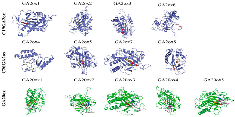
Figure 3.
3D structures of LcGA oxidases showing functional sites. The conserved motif related GA substrate binding site and 2-oxoglutarate-binding motif were display the purple and blue bands. The amino acid residues that bind the Fe2+ and interacted with the 5-carboxylate of 2-oxoglutarate are highlighted in yellow and red, respectively.

