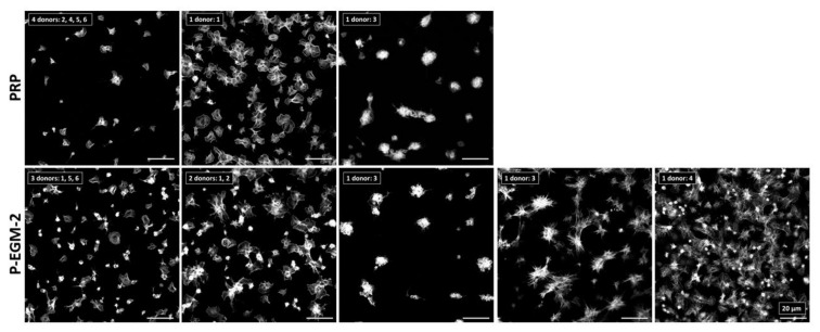
Figure 2.
Adherence of platelets (PLT) on poly(tetrafluoro ethylene). Platelet-rich plasma (PRP) was incubated under static conditions for 1 h at 37 °C with basal medium containing nine supplements (P-EGM-2) in a ratio of 1:1 resulting in a concentration of 50,000 PLT µL−1. PRP served as control. PLT of six different donors (numbered from 1–6) were stained with phalloidin conjugated with Alexa Fluor 555 and images were taken by confocal laser scan microscopy at 100-fold primary magnification. Shown are different classes of platelet morphologies (with the individual donors representing them as indicated) after incubation with the complete medium (P-EGM-2; 5 different classes) or without any medium components (PRP; 3 different classes). For example, PLT of four donors (No. 2, 4, 5 and 6) incubated with PRP adhered in low numbers and show a spread morphology. Scale bar represents 20 µm.

