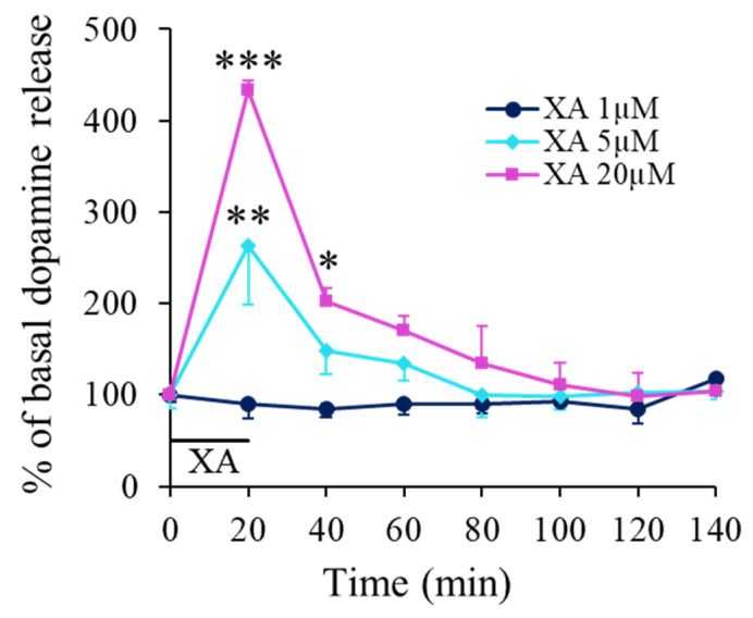
Figure 2.
XA-induced DA release in the prefrontal cortex. A. Time evolution of DA release measured by microdialysis. Filled symbols represent the mean percentage (±SEM) of the mean basal DA release (mean of 8 × 20 min dialysate fractions before retro-dialysis of XA) measured in consecutive 20 min cumulated dialysate. Continuous lines are interpolation between data points. The black bar indicates the period of XA application at concentration of 1, 5 or 20 µM as indicated. The dopamine basal release level (100%) was 433 ± 32 fmol/20 min for the 20 µM-treated group (n = 3 to 5 rats per group). Under the influence of XA administered directly to the brain tissue, we observed a graduated increase in the extracellular dopamine with a local four-fold increase by reference to controls for a concentration of XA of 20 µM. The local diffusion of XA into the brain through the probe was estimated in vitro around 18%. * p < 0.05; ** p < 0.01; *** p < 0.001, comparison with control before XA infusion. Not corrected from in vitro recovery.

