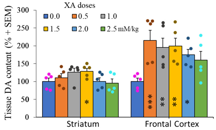
Figure 4.
Dose-effect of XA administration on dopamine concentrations in the frontal cortex and striatum. Histogram of mean (+SEM) DA tissue content as a function XA doses as indicated. Rats were sacrificed 60 min after i.p injection of XA. As previously described [25], the i.p. administration of 50 mg/kg of XA to the rats induced a strong increase in the brain concentration of this compound (frontal cortex and caudate putamen in particular). The figure shows a two-fold increase of DA concentrations in the frontal cortex after 0.5 mM/kg and these concentrations remain stable at higher doses of XA. By contrast, the change of DA-tissue content in the striatum is very low and almost not significant, even at the highest concentration of XA. Filled circles represent individual data point sets for each bar of the histogram accordingly. * p < 0.05; ** p < 0.01; *** p < 0.001; post ANOVA comparison with control animals using the uncorrected Fisher’s LSD statistical test.

