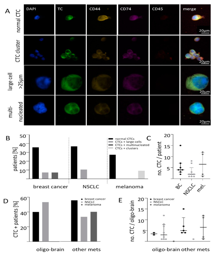
Figure 1.
Liquid biopsy analysis of brain metastatic patients. (A) Xcyto®10 analysis of liquid biopsies from patients with breast cancer, NSCLC and melanoma revealed different kind of CTCs enriched with Parsortix from 7.5 mL whole blood. (B) CTCs were detected in 50% of breast cancer, in 47% of NSCLC and in 36% of melanoma patients with mostly typical CTCs (black). The CTC positive patients were further subdivided as only normal CTCs, normal CTCs and large cells, normal CTCs and multinucleated cells as well as normal CTCs and clusters. Additionally, CTC clusters could be detected in the melanoma cohort (light grey) and some patients had tumor marker-positive large cells (mid grey) or multinucleated cells (dark grey). (C) No difference in the average number of CTCs between the different tumor entities. (D) Patient cohort divided by the metastatic spread shows varying CTC positivity. (E) No difference in the median CTC numbers could be observed in relation to the metastatic site (breast cancer p = 0.160; NSCLC, p = 0.419, melanoma p = 0.400).

