Figure 2.
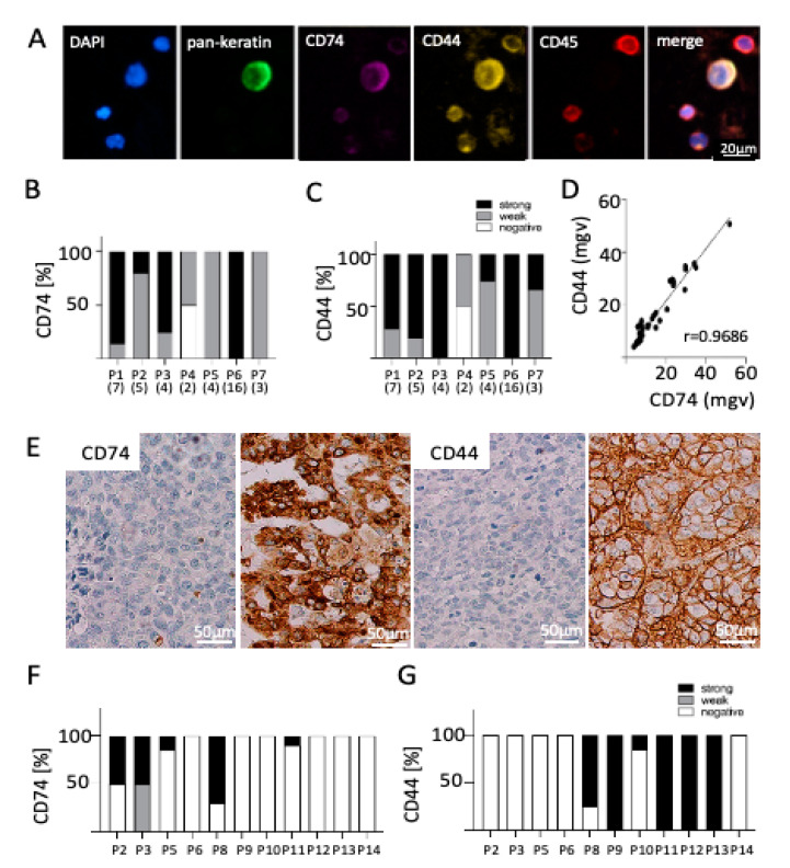
CD74 and CD44 expression on CTCs and matched BM in breast cancer. (A) Exemplary CTC positive for pan-keratin, CD74, and CD44 and negative for CD45 (leukocyte exclusion marker) surrounded by four leucocytes positive for CD45 and for both CD44 and CD74. Bar chart showing the (B) CD74 and (C) CD44 expression on CTCs in seven breast cancer BM patients. Number in parenthesis represents the number of CTCs identified in each patient. Most patients showed a weak or high expression of both proteins on their CTCs. (D) Correlation analysis revealed that CD74 and CD44 are expressed to a similar extent on enriched CTCs (r = 0.9686; mgv = mean grey value). (E) IHC analysis of matched brain metastasis revealed differential expression of (F) CD74 and (G) CD44 independent of CTC status (P2–P6: CTC-positive patients, P8-P14 CTC-negative patients), determined by H-score (see method section for details). Each bar indicates the expression pattern of one brain metastasis sample per patient.

